ULTRASOUNDS
AND PRENATAL TESTS
ULTRASOUND AND PRENATAL TESTING UNIT
The head and director of the unit is Dr. Gerard Albaigés, a gynecologist specializing in fetal medicine with exclusive training and dedication to this field of medicine.
He is currently the director of Fetal Medicine R&D at the Hospital Universitari Dexeus in Barcelona. He is also the coordinator of the ultrasound and fetal medicine section of the Catalan Society of Gynecology and Obstetrics and is a member of the Fetal Medicine Foundation based in London.

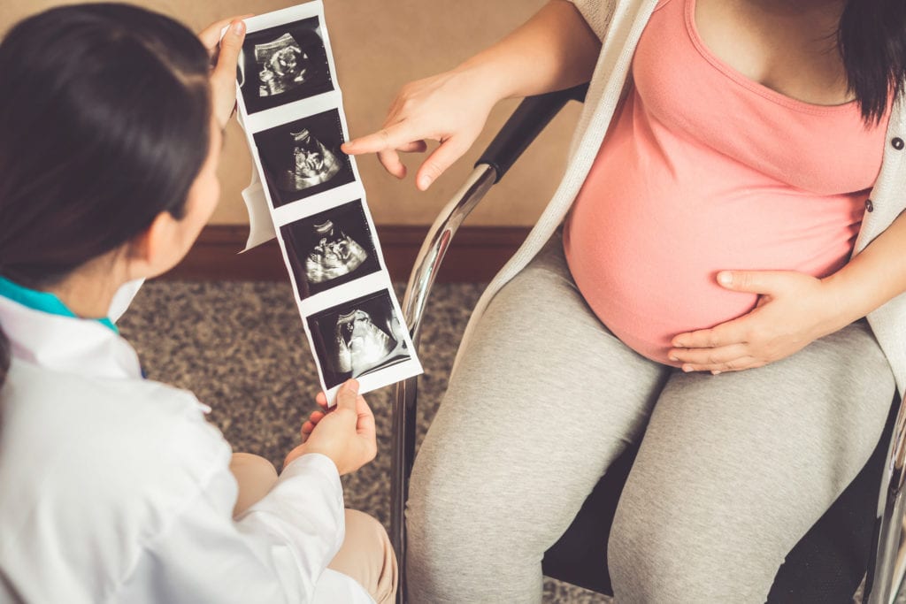
Confidence in the monitoring and control of your baby is essential to enjoy a healthy pregnancy.
For a peaceful pregnancy, get ready and get to know your child with the most modern prenatal diagnosis in the hands of the best professionals in the sector in ultrasound and prenatal tests.
PREGNANCY WEEK BY WEEK


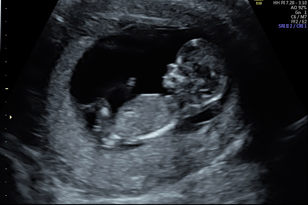
EARLY FIRST TRIMESTER ULTRASOUND AT 8 WEEKS OF GESTATION
Ultrasound performed between 7 and 9 weeks of gestation to check the number of embryos and the presence of a positive fetal heartbeat
SCREENING FOR RISK OF CHROMOSOMAL DISORDERS (DOWN SYNDROME)
It consists of a maternal blood test (week 9-11) to measure some hormones (B-hCG and PAPP-A protein) and a 12-week ultrasound where a fetal anatomical examination is performed. From the data obtained together with the maternal age, the risk of suffering chromosomal alterations in the fetus is calculated. The results obtained are explained in detail and accompanied by appropriate prenatal advice.
NON-INVASIVE CHROMOSOME ANALYSIS: TRISONIM NON-INVASIVE PRENATAL TEST (10-13 WEEKS)
The non-invasive prenatal test allows the detection of alterations in chromosomes 13, 18, 21 and also the determination of the fetal sex (X and Y chromosomes) as well as the detection of certain microdelections (loss of portions of the chromosomes). With a simple analysis of the mother, more than 98.5% of the results for the analysed chromosomes are detected. It can be applied both in single and double pregnancies, with the same reliability.
INVASIVE TECHNIQUES
They are performed transabdominally using local anesthesia. The risk of current fetal loss is 1:500. The main result is obtained in 3 days and the definitive one in 3 weeks.
Type of techniques:
-
- Chorionic biopsy (12-13 weeks)
A fetal karyotype is performed on the chorionic tissue obtained in the biopsy. It is especially useful in suspected early chromosomal disorders. The Embriogyn unit performs this test transabdominally.
-
- AmniocentesisAmniocentesis
A fetal karyotype is performed on the fetal cells obtained from the amniotic fluid. Most indications come from fetal structural alterations or maternal desire.

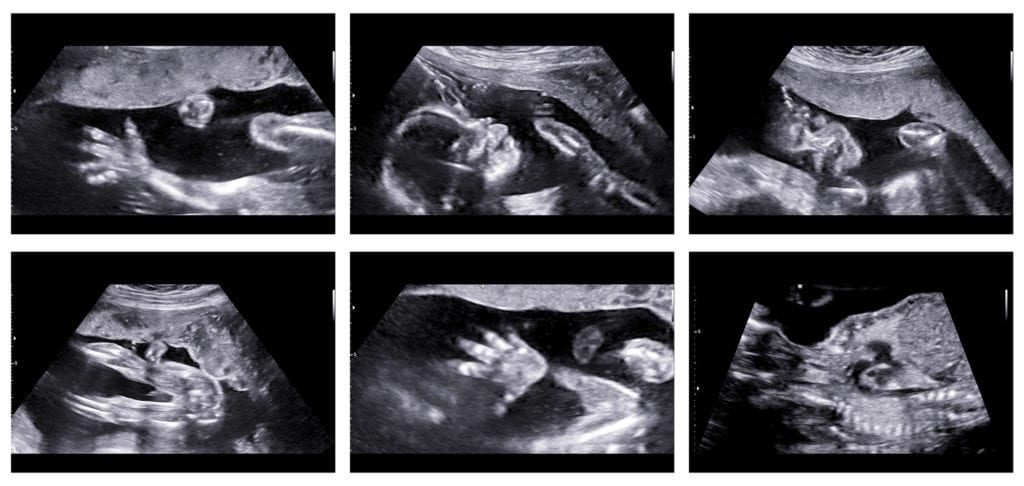
HIGH DEFINITION MORPHOLOGICAL ULTRASOUND EVALUATION
It is the so-called high definition or morphological ultrasound scan in which the fetal anatomy is evaluated in a systematized way, looking for different structural alterations in all the organs and skeleton for a correct planning of the pregnancy and delivery.
By means of color and pulsed Doppler, the resistance indexes in the uterine arteries are measured in order to define the risk factors of preeclampsia or intrauterine growth retardation.
4D-5D ULTRASOUND
FIVE-DIMENSIONAL ULTRASOUND ASSESSMENT
It is performed with state-of-the-art ultrasound technology. It is about obtaining spectacular images in volume and in real time, which allow parents to create a very strong mother-child bond, since it is anticipated and allows us to see how our child will be before birth.
The sharpness and resolution of the images in this technique provide greater diagnostic capacity for anomalies. It means a new generation and perspective in terms of the power of the images that provide an increase in resolution and diagnostic capacity.

BIOMETRICS: THIRD QUARTER ULTRASOUND ASSESSMENT
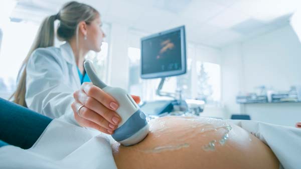
It is performed by an ultra-fast ultrasound machine with excellent resolution for obstetric examinations. The anatomy is analysed and examined for abnormalities that did not appear on previous ultrasound scans. Fetal biometry is used to determine the state of fetal growth and to inform the patient of possible risks that may arise during the third trimester. The placenta is assessed for ultrasound appearance and position. From this third trimester ultrasound scan, the type and specifications of the delivery are planned.
MAKE AN APPOINTMENT
It’s quite simple. All you have to do is contact Embriogyn and make an appointment with our specialists at the time that suits you best. In case you can’t come in person to the clinic, visits can also be arranged via Skype.
